If your doctor has recommended imaging, you may be wondering about the difference between a PET scan and a CT scan, and which one is right for your situation. Both are powerful diagnostic tools used every day in medicine, but they work in fundamentally different ways and provide different types of information.
The simplest way to think about it:
-
- A CT scan shows structure and anatomy — what your organs, bones, and tissues look like.
-
- A PET scan shows function and metabolic activity — how your cells are behaving at a biological level.
Understanding these differences can help you feel more informed and prepared when your physician orders imaging. In this guide, we break down how each scan works, when each is used, and how they compare, especially when it comes to cancer detection.
What Is a CT Scan?
A CT scan (computed tomography scan) uses a series of X-ray images taken from different angles around the body, which a computer then combines to create detailed cross-sectional images. These images allow physicians to see bones, organs, blood vessels, and soft tissues in high resolution which can be far more detailed than a standard X-ray provides.
CT scans are one of the most commonly performed imaging studies in medicine. They are fast (most scans take 10 to 30 minutes), widely available, and provide excellent structural detail. A contrast dye may be injected intravenously or taken orally before the scan to help certain tissues and blood vessels show up more clearly.
CT scans are commonly used to:
-
- Detect and locate tumors, masses, or abnormal growths
-
- Evaluate injuries to bones, organs, and internal structures
-
- Guide surgical planning and biopsy procedures
-
- Monitor treatment response by measuring changes in tumor size
-
- Diagnose conditions affecting the lungs, abdomen, pelvis, and brain
What Is a PET Scan?
A PET scan (positron emission tomography) works differently. Rather than imaging the body’s structure, a PET scan reveals how cells are functioning at the metabolic level. Before the scan, a small amount of a radioactive tracer, most commonly fluorodeoxyglucose (FDG), a form of sugar, is injected into the bloodstream. Because cancer cells typically consume glucose at a much higher rate than normal cells, the tracer accumulates in areas of high metabolic activity. The PET scanner then detects this concentration and creates a three-dimensional map showing where that activity is occurring.
PET scans are particularly valuable in oncology because they can detect cancer activity before structural changes are visible on a CT scan. A tumor may be too small to appear on a CT image, but its elevated metabolic activity can already be detected on a PET scan.
PET scans are commonly used to:
-
- Detect cancer and determine whether it has spread (metastasized)
-
- Differentiate between benign and malignant growths
-
- Evaluate how well a cancer is responding to treatment
-
- Identify cancer recurrence after treatment
-
- Assess metabolic activity in the brain and heart in certain conditions
Key Differences Between PET Scan and CT Scan
Difference Between PET Scan and CT Scan
| Feature | CT Scan | PET Scan |
|---|---|---|
| What It Shows | Structure and anatomy | Metabolic function and cellular activity |
| How It Works | X-ray beams from multiple angles | Radioactive tracer (FDG) uptake |
| Best For | Detecting size, shape, and location of abnormalities | Detecting cancer activity, spread, and treatment response |
| Imaging Type | Structural (anatomical) | Functional (cellular/metabolic) |
| Scan Duration | 10–30 minutes | 60–90 minutes (including tracer uptake time) |
| Radiation Source | External X-rays | Injected radioactive tracer |
| Contrast Agent | Iodine-based IV contrast (optional) | FDG tracer (required) |
| Resolution | High structural detail | Lower structural detail, high metabolic detail |
Beyond these core differences, the two scans also differ in how quickly results are available. CT scans typically produce results within hours, while PET scan interpretation may take one to two days due to the complexity of metabolic analysis. Cost also differs between the two as PET scans are generally more expensive than CT scans, though both are typically covered by insurance when medically indicated.
What Is a PET/CT Scan?
A PET/CT scan combines both technologies into a single examination performed on one machine. The patient undergoes both scans during the same session because the PET component captures metabolic activity while the CT component provides the structural roadmap. The two image sets are then fused together by computer software, creating a combined image that shows exactly where abnormal metabolic activity is occurring within the body’s anatomy.
This combination is more powerful than either scan alone. A PET scan might show a “hot spot” of metabolic activity, but without the CT overlay, it can be difficult to pinpoint the exact anatomical location. Conversely, a CT scan might reveal a mass, but cannot tell you whether that mass is metabolically active (potentially cancerous) or inactive (potentially benign).
PET/CT has become the standard of care in oncology for cancer staging, treatment planning, and monitoring. At Ackerman Cancer Center, PET/CT imaging is performed using advanced scanners that deliver precise, high-resolution combined images, giving our radiation oncologists the detailed information they need to design accurate, targeted treatment plans.
PET Scan vs CT Scan for Cancer
CT Scan vs PET Scan for Cancer
When it comes to cancer, CT scans and PET scans each play critical but different roles:
-
- CT scans for cancer excel at showing the size, shape, and exact location of tumors. They are often the first imaging study ordered when cancer is suspected, and they are essential for surgical planning and measuring whether tumors are shrinking during treatment. CT scans can detect tumors throughout the body including lungs, liver, abdomen, pelvis, brain, and more.
-
- PET scans for cancer reveal whether cells are behaving like cancer cells and consuming glucose at abnormally high rates. This is critical for determining whether a tumor is malignant, whether cancer has spread to lymph nodes or distant organs, and whether a residual mass after treatment contains active cancer or is just scar tissue.
Which Is Better?
For cancer detection and management, PET and CT scans are most effective when used together as a PET/CT scan. The combined study provides both the metabolic and structural information that oncologists need to make treatment decisions with confidence. Which individual scan is “better” depends entirely on the clinical question being asked:
-
- Need to know the size and location of a known tumor? CT scan.
-
- Need to know whether cancer has spread or whether treatment is working? PET scan.
-
- Need the most complete picture for staging, planning, or monitoring? PET/CT scan.
PET Scan vs CAT Scan — Is There a Difference?
This is one of the most common questions patients ask, and the answer is simple: a CAT scan and a CT scan are the same thing. “CAT scan” stands for “computed axial tomography,” while “CT scan” stands for “computed tomography.” The medical community shifted to the shorter “CT scan” terminology over time, but both terms refer to the identical procedure using the same technology. If your doctor mentions a CAT scan, they are ordering a CT scan.
When Do You Need a PET Scan vs CT Scan?
Your Radiation Oncologist determines which scan to order based on the specific clinical question:
-
- CT scan is typically ordered when your doctor needs to evaluate an injury, locate a mass, check for internal bleeding, assess organ health, or plan a surgical procedure. CT scans are also the standard follow-up imaging for monitoring tumor size during and after cancer treatment.
-
- PET scan is typically ordered when your doctor needs to determine whether a mass is cancerous, evaluate whether cancer has spread (staging), assess treatment response at the cellular level, or check for cancer recurrence. PET scans are most commonly ordered by oncologists for patients with known or suspected cancer.
-
- PET/CT scan is ordered when both metabolic and structural information are needed, which is the case for most cancer staging and treatment planning decisions.
In many cases, you may receive a CT scan first, and if the results raise questions about cancer activity, a PET scan or PET/CT scan will follow to provide more complete information.
Benefits and Limitations of Each Scan
CT Scan Pros and Cons
Benefits:
-
- Fast — most scans completed in 10 to 30 minutes
-
- Widely available at hospitals and imaging centers
-
- Excellent structural detail for bones, organs, and soft tissues
-
- Lower cost compared to PET scans
-
- Effective for emergency and trauma evaluation
Limitations:
-
- Cannot distinguish between benign and malignant masses based on appearance alone
-
- Uses ionizing radiation (though doses are low and carefully controlled)
-
- Contrast dye may cause allergic reactions or kidney concerns in some patients
-
- May miss small or early-stage cancers that have not yet formed detectable masses
PET Scan Pros and Cons
Benefits:
-
- Detects cancer activity at the cellular level, often before structural changes appear
-
- Can differentiate between active cancer and scar tissue or benign masses
-
- Provides whole-body assessment in a single scan
-
- Critical for accurate cancer staging and treatment response evaluation
Limitations:
-
- More expensive than CT scans
-
- Longer procedure time (60 to 90 minutes including tracer uptake)
-
- Lower structural resolution; anatomical detail is less precise
-
- Some cancers (slow-growing tumors, certain types of prostate cancer) may not show high FDG uptake
-
- Requires fasting before the scan; blood sugar levels must be controlled
What to Expect During Each Scan
CT Scan Procedure
-
- You may be asked to change into a hospital gown and remove metal jewelry or accessories
-
- If contrast is needed, an IV line will be placed; oral contrast may be given to drink beforehand
-
- You lie on a table that slides into the CT scanner; a large, ring-shaped machine
-
- The scan itself takes 10 to 30 minutes; you will need to lie still and may be asked to hold your breath briefly
-
- The procedure is painless; you may feel warmth from the contrast injection
PET Scan Procedure
-
- You will fast for 4 to 6 hours before the scan (water is allowed)
-
- The FDG radioactive tracer is injected through an IV; this is painless
-
- You rest quietly for 45 to 60 minutes while the tracer circulates and is absorbed by metabolically active cells
-
- You then lie on a table that moves through the PET scanner for approximately 20 to 30 minutes
-
- The entire appointment takes approximately 2 to 3 hours from check-in to completion
For PET/CT scans, both procedures are performed during the same session, the PET and CT images are acquired sequentially on a combined scanner, so you do not need to schedule separate appointments.
PET vs CT Scan — Which Is Right for You?
The right scan depends on your individual medical situation and what your physician needs to learn. In many oncology cases, both scans will be part of your diagnostic and treatment journey. Rather than choosing one over the other, the goal is to use the right imaging tool at the right time to get the most accurate and actionable information.
If you have questions about which imaging studies are appropriate for your situation, a consultation with a specialist can help you understand your options and what each scan will reveal. At Ackerman Cancer Center, our team works closely with referring physicians to ensure patients receive the imaging studies that provide the clearest, most complete diagnostic picture.
Where to Get PET and CT Scans in Northeast Florida
Ackerman Cancer Center provides advanced diagnostic imaging services at our Northeast Florida locations:
-
- Mandarin (Jacksonville, FL)
-
- Amelia Island, FL
-
- St. Augustine, FL (World Golf Village area)
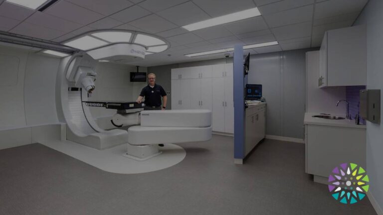
Our on-site imaging state-of-the-art PET/CT scanners and advanced CT technology, operated by experienced radiology professionals. As the world’s first private physician-owned proton therapy practice, Ackerman Cancer Center brings the same precision-focused approach to diagnostic imaging that has guided our treatment philosophy since 1997. Our board-certified specialists review every scan to ensure accurate interpretation and seamless coordination with your treatment team.
Frequently Asked Questions (FAQ)
What is the difference between a PET scan and a CT scan?
A CT scan uses X-rays to create detailed images of the body’s structure showing the size, shape, and location of organs, bones, and abnormalities. A PET scan uses a radioactive tracer (FDG) to detect metabolic activity at the cellular level, revealing how cells are functioning rather than just what they look like. CT scans show anatomy; PET scans show biology.
Is a PET scan better than a CT scan?
Neither is universally “better”, rather they answer different questions. CT scans are better for evaluating structural detail, injuries, and tumor size. PET scans are better for detecting cancer activity, staging, and treatment response. For cancer care, the two scans are most powerful when combined as a PET/CT scan, which provides both structural and metabolic information in one study.
Why would a doctor order a PET scan instead of a CT scan?
A doctor typically orders a PET scan when they need to evaluate metabolic activity. For example, to determine whether a mass is cancerous, whether cancer has spread to other parts of the body, whether a treatment is working at the cellular level, or whether a residual mass after treatment contains active cancer or scar tissue. These are questions that a CT scan alone cannot answer.
Can a CT scan detect cancer as well as a PET scan?
CT scans can detect tumors and masses based on their size and appearance, but they cannot determine whether a mass is metabolically active (potentially cancerous) or inactive (potentially benign). Small or early-stage cancers that have not yet formed a visible mass may be missed on CT. PET scans detect cancer based on cellular behavior, which can identify malignancies at earlier stages and in locations that CT may not reveal. However, CT scans provide superior structural detail that PET scans lack, which is why the combination (PET/CT) is the gold standard.
What is a PET/CT scan and why is it used?
A PET/CT scan combines both imaging technologies into a single examination. The PET component shows metabolic activity, while the CT component provides an anatomical roadmap. The images are fused together by computer, showing exactly where abnormal cellular activity is occurring within the body. PET/CT is the standard of care for cancer staging, treatment planning, and monitoring because it provides the most complete diagnostic picture available.
Which scan is more accurate for cancer detection?
For detecting the presence and spread of cancer, PET/CT is generally the most accurate option because it combines metabolic and structural data. A PET scan alone is highly sensitive to cancer activity but has lower anatomical resolution. A CT scan alone provides excellent structural detail but cannot assess whether a mass is biologically active. When accuracy matters most, as it does in cancer staging and treatment decisions, PET/CT provides the most reliable and comprehensive information.
Are PET scans or CT scans safer?
Both scans involve exposure to radiation, and both are considered safe when performed for appropriate medical indications. CT scans use external X-rays, while PET scans use an internally injected radioactive tracer that naturally decays and is eliminated from the body within hours. The radiation doses from both scans are carefully controlled and are low relative to the diagnostic value they provide. Your physician weighs the benefits of the information gained against the minimal risks before ordering any imaging study.
How long does each scan take?
A CT scan typically takes 10 to 30 minutes. A PET scan takes longer, approximately 2 to 3 hours total, including 45 to 60 minutes of quiet rest while the tracer is absorbed, followed by 20 to 30 minutes of scanning. A combined PET/CT scan is performed in a single session and takes approximately 2 to 3 hours overall.
Do I need both scans?
In oncology, yes, it is common to need both. Many patients receive a CT scan as an initial diagnostic study, followed by a PET scan or PET/CT scan for more detailed cancer staging and treatment planning. During and after treatment, both imaging types may be used at different points to monitor progress. Your oncologist will determine which imaging studies are appropriate based on your specific diagnosis and treatment plan.
Where can I get a PET or CT scan in Northeast Florida?
Ackerman Cancer Center offers advanced PET/CT and CT imaging at our locations in Mandarin (Jacksonville), Amelia Island, and St. Augustine (World Golf Village area). Our board-certified specialists and experienced imaging team provide accurate, timely diagnostic imaging with seamless coordination for patients undergoing cancer evaluation or treatment. Call (904) 880-5522 or schedule an appointment to learn more.
Contact Ackerman Cancer Center Today
Getting the right diagnostic imaging is a critical first step in understanding your health and making informed treatment decisions. Whether you need a CT scan, PET scan, or PET/CT scan, Ackerman Cancer Center provides advanced imaging technology and expert interpretation at convenient Northeast Florida locations.
-
- Schedule an imaging appointment — our team will coordinate with your referring physician
-
- Talk to a specialist about which imaging studies are right for your situation
Verify your insurance — call (904) 880-5522 to confirm coverage before your visit