Deciding when and how to screen for breast cancer can feel confusing, especially when you hear terms like 3D mammography or contrast-enhanced mammography. If you are looking for clear guidance on breast cancer screening in Northeast Florida, this guide breaks down each option, who it is for, and how to choose. It is written for women navigating screening decisions, particularly those with dense breast tissue or for those at a higher-than-average risk of developing breast cancer.
Early detection remains one of the most powerful tools against breast cancer. Below, we cover current screening guidelines, explain breast density in plain language, compare mammogram types, and show why Ackerman Cancer Center is leading in medical advancements as the first and only provider of contrast-enhanced mammography in Northeast Florida.
Key Takeaways
- The American Cancer Society notes women have the option to begin annual mammograms at age 40, with annual screening recommended from 45.
- About 40% of women have dense breast tissue, which can make cancers harder to see on a standard mammogram.
- 3D mammography (tomosynthesis) captures layered images and improves detection in many patients.
- Contrast-enhanced mammography (CEM) uses a contrast agent to highlight areas of concern, especially useful for dense breasts and higher-risk patients.
- Ackerman Cancer Center is the first and only CEM provider in Northeast Florida.
When to Start Breast Cancer Screening
Screening guidelines have evolved, and the right starting age can depend on your personal risk. The American Cancer Society recommends that women with average risk have the option to begin annual mammograms at age 40, should begin annual screening by age 45, and can transition to every two years at age 55 if they choose, continuing as long as they are in good health.
Women at higher risk, such as those with a strong family history or a known genetic mutation like BRCA, may need to start earlier and use additional imaging. The American College of Radiology recommends that all women have a formal risk assessment by age 25 to identify those who would benefit from earlier or supplemental screening.
Know Your Personal Risk
Risk factors include family history, genetic mutations, prior chest radiation, and dense breasts. A conversation with your physician about your personal risk is the best way to set a screening plan. If you are at higher risk, the screening plan may include imaging beyond a standard annual mammogram.
Understanding Breast Density
Breast density is one of the most important, yet, least understood parts of screening. It has nothing to do with how your breasts feel and everything to do with how they look on a mammogram.
What Dense Breasts Mean
Breasts are made of fatty tissue and dense glandular and fibrous tissue. On a mammogram, fat looks dark while dense tissue looks white. The problem is that cancer also looks white, so dense tissue can hide a tumor, like trying to spot a snowflake in a snowstorm.
Roughly 40% of women have dense breasts. Because density can mask cancers and is itself a modest risk factor, many states now require that mammogram reports notify women about their breast density. The American College of Radiology supports informing patients so they can discuss supplemental screening options.
Why This Matters for Your Screening Choice
If you have dense breasts, a standard mammogram alone may not be enough. This is where advanced options like contrast-enhanced mammography become especially valuable, and why a center offering multiple screening tools can better personalize your care.
Comparing Your Screening Options
Not all mammograms are the same, and newer tools add detection power for the right patients. Here is how the main options compare.
2D Mammography
A traditional mammogram takes two X-ray images of each breast. It is fast, widely available, and remains the foundation of breast cancer screening. For women with mostly fatty breast tissue, it works very well.
3D Mammography (Tomosynthesis)
3D mammography, also called tomosynthesis, captures multiple thin image layers of the breast, which the radiologist can scroll through, like pages in a book. This reduces the “overlap” problem of 2D imaging and can improve cancer detection while lowering the number of callbacks for false alarms.
Contrast-Enhanced Mammography (CEM)
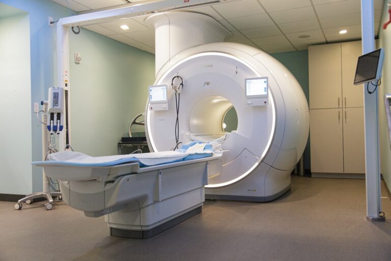
Contrast-enhanced mammography is an advanced breast imaging exam that combines a digital mammogram with an iodine-based contrast agent given through an IV. Because many breast cancers develop increased blood flow, the contrast helps highlight suspicious areas that may be difficult to see on a standard mammogram, especially in women with dense breast tissue.
CEM provides both anatomical and functional imaging in a single exam, making it valuable for evaluating abnormal findings, assessing the extent of newly diagnosed breast cancer, and screening select women at higher risk.
Ackerman Cancer Center is the first and only provider of CEM in Northeast Florida, offered through the Ackerman Breast Clinic – Amelia Island.
Screening Comparison Table
| Option | Best For | How It Works | Key Advantage |
|---|---|---|---|
| 2D mammogram | Average risk, fatty tissue | Two X-ray views | Fast, foundational |
| 3D mammography | Most patients | Layered image slices | Better detection, fewer callbacks |
| CEM | Dense breasts, higher risk | Mammogram plus IV contrast | Highlights cancers by blood flow |
What to Expect and How to Prepare for a Mammogram
Knowing what happens can ease anxiety. A standard mammogram takes about 20 minutes, and you will stand while each breast is briefly compressed between two plates for the images. Compression can feel uncomfortable but lasts only seconds per view.
Preparing for Your Appointment
For all mammograms:
- Schedule your exam for the week after your period, if applicable, when breasts are often less tender.
- Do not wear deodorant, powder, lotion, or perfume on your underarms or breasts on the day of your exam, as these can appear on the images.
- If you are a new patient, arrange to have prior breast imaging sent to us prior to your appointment so your radiologist can compare your current exam with previous studies.
For Contrast-Enhanced Mammography (CEM):
- You will receive an iodine-based contrast agent through an IV before your exam.
- Let your care team know if you have a history of contrast allergies, kidney disease, or if you are pregnant or think you may be pregnant.
- Drink plenty of water before and after your appointment unless your provider has instructed otherwise.
- Your appointment will take slightly longer, approximately 45 minutes, to allow time for the IV and contrast administration.
After Your Screening
The staff at Ackerman Cancer Center evaluate the images and notify patients once the image results are finalized. If something needs a closer look, the on-site diagnostics teams at Ackerman Cancer Center, including breast ultrasound and biopsies, mean follow-up can often happen quickly rather than after weeks of waiting. Explore all options on our screening and diagnostic services page, and learn about treatment on our breast cancer page.
Frequently Asked Questions
At what age should I start my annual mammogram?
The American Cancer Society recommends that women at average risk can begin annual mammograms at age 40 and should begin annual screening by 45. Women at higher risk may need to begin earlier screenings. Talk with your physician about your personal risk to set the right schedule for you.
What is the difference between a mammogram and CEM?
A standard mammogram uses X-rays to image the breast. Contrast-enhanced mammography (CEM) adds an iodine-based contrast agent through an IV that highlights areas with increased blood flow, which can reveal cancers hidden in dense breast tissue. CEM is especially helpful for dense breasts and higher-risk patients.
What does it mean to have dense breasts?
Dense breasts have more glandular and fibrous tissue relative to fat. On a mammogram, both dense tissue and cancer appear white, so density can hide tumors. About 40% of women have dense breasts. If you do, your physician may recommend supplemental screening like Contrast Enhanced Mammography (CEM)
Is contrast-enhanced mammography available in Northeast Florida?
Yes. Ackerman Cancer Center is the first and only provider of contrast-enhanced mammography (CEM) in Northeast Florida, offered through the Ackerman Breast Clinic – Amelia Island. It is particularly valuable for women with dense breasts or elevated risk. Contact the clinic to learn whether CEM is appropriate for you.
Does a mammogram hurt?
No. A mammogram involves brief compression of the breast, which can feel uncomfortable but usually lasts only seconds per image. The full exam takes about 20 minutes. Scheduling during the week after your period, when applicable, can reduce tenderness and make the experience more comfortable.
Schedule Your Screening With Confidence
Breast cancer screening in Jacksonville is not one-size-fits-all. Whether a standard mammogram, 3D mammography, or contrast-enhanced mammography is right for you depends on your age, risk, and breast density. The key is having access to every option and a team that helps you choose wisely, which is exactly what the Ackerman Breast Clinic provides as Northeast Florida’s first and only CEM provider.
Early detection saves lives, and the best screening is the one you actually schedule. Contact us today or visit the Ackerman Breast Clinic to book your appointment. We are here to support you every step of the way.
Medical disclaimer: This article is for general educational purposes and is not medical advice. Screening recommendations vary by individual risk and should be made with a qualified physician. This content does not replace a personal consultation or professional evaluation of any symptom or finding.